Cell: The Building Block of Life Class 9 Notes: Looking for complete and easy-to-understand Class 9 Science Chapter 2 notes? You are at the right place. In this post, we have provided comprehensive notes for Cell: The Building Block of Life from the CBSE Class 9 Science Exploration textbook. These notes cover all the important concepts of the chapter in simple language, with clear definitions, key points, comparisons, important terms, and well-labelled diagrams to make learning and revision easier. The notes are designed to help students understand concepts quickly, prepare for school examinations, and revise the chapter effectively. Students can also download the complete Class 9 Science Chapter 2 notes in PDF format using the download link provided below.
Table of Contents
The organisms found in these hot springs are mostly thermophiles.
- Thermophiles are heat-loving bacteria.
- They are unicellular organisms.
Cell — The Basic Unit of Life
All living organisms are made up of cells. The cell represents the basic level at which life exists. Organisms can be:
- Unicellular: made up of a single cell.
- Examples: bacteria and yeast.
- Multicellular: made up of millions of cells that work together.
- Examples: plants, fish, birds and humans.
The cell is called the structural and functional unit of life because:
- All living organisms are made up of one or more cells.
- A cell forms the basic structure of an organism.
- Cells perform essential life processes such as obtaining energy, synthesising substances, removing waste and reproducing.
- In multicellular organisms, cells are organised in a hierarchy:
- Cells → Tissues → Organs → Organ Systems → Organism
How to Study Cells?
1. Limit of Resolution
Limit of resolution is the ability of the human eye to see two very close objects as separate and distinct.
- At a viewing distance of about 25 cm, the human eye can distinguish two points separated by about 0.1 mm.
- Therefore, the limit of resolution of the human eye is 0.1 mm.
2. Magnification
Magnification is the process of making an object appear larger so that its details can be observed.
- A convex lens or a combination of lenses can be used for magnification.
- In a microscope, the objective lens and eyepiece together magnify the object.
3. Robert Hooke and the Discovery of Cells
- Robert Hooke was the first person to observe cells in 1665.
- He used a self-designed microscope capable of about 200–300× magnification.
4. Light Microscope
A light microscope uses visible light and lenses to produce a magnified image.
In school laboratories, light microscopes are commonly used to observe cells and other fine structures.
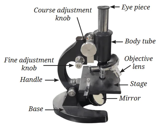
Important features of a good microscope
Scientists have improved microscopes mainly by improving:
- Resolution — clarity with which two close objects can be distinguished.
- Contrast — difference in brightness between different parts of an image.
- Magnification — ability to make the object appear larger.
| Estimated size of one cell=Number of cells along the diameter/Diameter of visible field in µm |
1 millimetre (mm) = 1000 micrometres (µm)
5. Total Magnification of a Microscope
The total magnification depends on the magnifying powers of the eyepiece and objective lens.
Formula
| Total magnification = Eyepiece magnification × Objective magnification |
Apart from light microscopes, scientists use powerful electron microscopes to study very fine cellular structures.
Light Microscope vs Electron Microscope
| Light Microscope | Electron Microscope |
|---|---|
| Uses visible light | Uses a beam of electrons |
| Used commonly in school laboratories | Used for detailed scientific studies |
| Provides lower level of detail | Reveals much finer details |
| Can study cells at the micrometre (µm) scale | Can reveal structures at the nanometre (nm) scale |
Structure of a Cell
The cell is the basic structural and functional unit of life. To function properly, a cell must interact with its surroundings and exchange materials with them. These activities are controlled mainly by the cell membrane, while the cell wall, cytoplasm, nucleus and cell organelles perform other important functions.
1. Cell Membrane — The Universal Feature of a Cell
The cell membrane, also called the plasma membrane, is a thin boundary that surrounds the cell and protects its contents.
Main functions
- It protects the contents of the cell.
- It defines the individuality of the cell.
- It controls the movement of substances into and out of the cell.
- It is selectively permeable, meaning it allows some substances to pass through while preventing others from passing through.
Structure of the Cell Membrane: The cell membrane is:
- Extremely thin — about 7–10 nm thick.
- Made mainly of lipids and proteins.
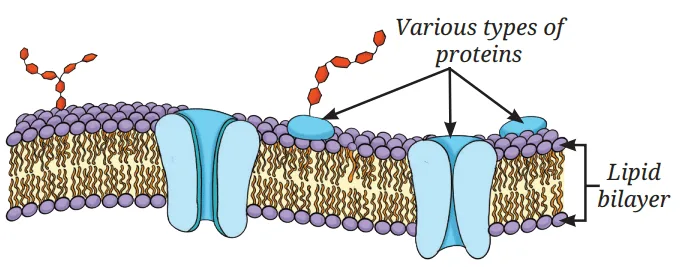
Fluid-Mosaic Model: According to this model:
- The membrane has a lipid bilayer, i.e. two layers of special lipid molecules.
- The water-attracting heads face outwards.
- The water-repelling tails face inwards.
- Various proteins are embedded in the lipid bilayer.
- Lipid and protein molecules can move sideways, flip and rotate; therefore, the membrane is called fluid.
- The arrangement of different molecules resembles a mosaic, giving rise to the term fluid-mosaic model.
- Membrane proteins act as gatekeepers, helping substances pass through the membrane.
2. Diffusion
Diffusion is the movement of particles from a region of higher concentration to a region of lower concentration.
- It occurs because of a concentration gradient.
- It can occur even without a membrane.
- Example: spreading of fragrance in air.
3. Osmosis
Osmosis is the movement of water through a selectively permeable membrane.
Water moves from a region having: More water + less solute → Less water + more solute
Thus, osmosis is a special type of diffusion involving water.
Example: In plants, water from the soil enters root cells through osmosis.
Diffusion vs Osmosis
| Diffusion | Osmosis |
|---|---|
| Movement of particles | Movement of water |
| From higher to lower concentration | Water moves through a selectively permeable membrane |
| Can occur without a membrane | Requires a selectively permeable membrane |
| Example: spreading of fragrance in air. | Example: In plants, water from the soil enters root cells through osmosis. |
4. Effect of Different Solutions on a Cell
The effect of a solution on a cell depends on its solute concentration compared with the cell.
(a) Isotonic Solution
- Solute concentration outside the cell = solute concentration inside the cell.
- There is no net movement of water.
- The cell remains approximately unchanged.
(b) Hypotonic Solution
- Solute concentration outside the cell is less than inside the cell.
- Water moves into the cell.
- The cell swells.
(c) Hypertonic Solution
- Solute concentration outside the cell is greater than inside the cell.
- Water moves out of the cell.
- The cell shrinks.
5. Cell Wall — The Outer Covering of Cells
Plants, fungi and bacteria have an additional covering outside the cell membrane called the cell wall.
Functions of the Cell Wall
- Provides rigidity and structural support.
- Protects the cell.
- Helps leaves and flowers remain firm.
- Helps plants maintain their shape.
- Helps plants stay upright.
- It is permeable, so water and some dissolved minerals can pass through it.
- Along with the selectively permeable cell membrane, its permeability helps plant roots absorb water and nutrients from the soil.
Composition: The plant cell wall is primarily made of cellulose.
Cell Wall and Osmosis
When a plant cell is placed in a concentrated sugar solution:
- Water moves out of the cell by osmosis.
- The inner contents shrink.
- However, the rigid cell wall maintains the overall shape of the cell.
- The cell membrane may pull away from the cell wall.
In contrast, animal cells do not have a cell wall, so they can shrink considerably when they lose water.
6. Cytoplasm
The cytoplasm is a semi-fluid, jelly-like substance present inside the cell.
It contains:
- the nucleus in eukaryotic cells, and
- various cell organelles and other substances.
Cell organelles perform specialised functions necessary for the survival and functioning of the cell.
7. Nucleus
The nucleus contains the cell's genetic information.
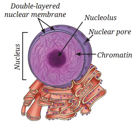
Structure of the Nucleus: The nucleus has:
- Nuclear membrane
- Double-layered covering.
- Contains pores through which materials can move between the nucleus and cytoplasm.
- Nucleolus
- A dense, round structure inside the nucleus.
- Ribosomal subunits are synthesised here.
- Chromatin
- Thread-like genetic material present in a non-dividing cell.
- Chromosomes
- Form when chromatin becomes organised as the cell prepares to divide.
- Contain DNA and specific proteins.
- DNA
- Contains the genetic information of the cell.
- Genes
- Functional segments of DNA that carry genetic information.
Simple sequence: Nucleus → Chromosomes → DNA → Genes
8. Prokaryotic Cells
Bacterial cells do not have a well-defined nucleus.
Instead:
- Their genetic material is present in a region called the nucleoid.
- The genetic material is not enclosed by a nuclear membrane.
- They also lack membrane-bound organelles.
Such cells are called prokaryotic cells.
Plant and animal cells have a well-defined nucleus and membrane-bound organelles and are therefore eukaryotic cells.
Comparison of Bacterial Cell vs Plant Cell vs Animal Cell Based on Their Structure
| Cell structures | Bacterial cell | Plant cell | Animal cell |
|---|---|---|---|
| 1. Cell membrane | ✓ Present | ✓ Present | ✓ Present |
| 2. Cell wall | ✓ Present | ✓ Present | ✗ Absent |
| 3. Cytoplasm | ✓ Present | ✓ Present | ✓ Present |
| 4. Well-defined nucleus (genetic material enclosed by a membrane) | ✗ Absent | ✓ Present | ✓ Present |
| 5. Primitive nucleus or nucleoid (genetic material without membrane around it) | ✓ Present | ✗ Absent | ✗ Absent |
| 6. Membrane-bound organelles | ✗ Absent | ✓ Present | ✓ Present |
Comparison between prokaryotic and eukaryotic cells
| Characteristics | Prokaryotic cell | Eukaryotic cell |
| 1. Primitive nucleus | Present | Absent |
| 2. Diameter of a typical cell | 1 to 10 µm | 10 to 100 µm |
| 3. Number of cells in an organism | Usually unicellular | Can be unicellular or multicellular |
| 4. Membrane-bound organelles | Absent | Present |
| 5. Membrane-bound nucleus | Absent | Present |
9. Cell Organelles
The cytoplasm of eukaryotic cells contains several specialised structures called cell organelles.
Each organelle performs a specific function. Together, they allow the cell to:
- build new materials,
- remove waste,
- produce energy, and
- carry out other life processes.
Important Cell Organelles and Their Functions
1. Ribosomes
- Ribosomes are tiny structures.
- They may be present freely in the cytoplasm or attached to the endoplasmic reticulum.
- They are the sites of protein synthesis.
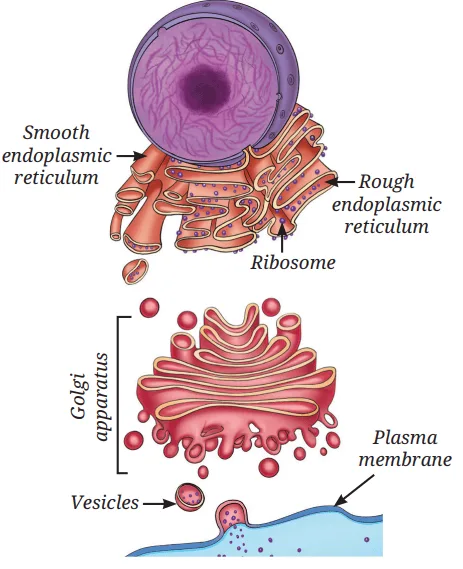
2. Endoplasmic Reticulum (ER)
- ER is a network spread throughout the cytoplasm.
- It is continuous with the outer membrane of the nuclear envelope.
- It helps in the synthesis and transport of proteins and lipids.
Rough Endoplasmic Reticulum (RER)
- Has ribosomes attached to its surface.
- Looks rough under an electron microscope.
- Mainly involved in protein synthesis and protein secretion.
Smooth Endoplasmic Reticulum (SER)
- Has no ribosomes attached.
- Looks smooth.
- Involved in the synthesis and storage of fats and hormones.
3. Golgi Apparatus
- Made up of stacks of flattened, sac-like structures.
- It is functionally linked with the ER, cell membrane and other organelles.
- It:
- modifies proteins and lipids,
- sorts them,
- packages them into vesicles,
- helps in their transport and secretion,
- and contributes to lysosome formation.
4. Lysosomes
- Lysosomes are single-membrane-bound sacs.
- They contain enzymes.
- These enzymes break down:
- unwanted proteins,
- carbohydrates,
- fats, and
- damaged or worn-out cell parts.
- The products of breakdown may be released into the cytoplasm and reused.
- Lysosomes are also known as the suicide bag (or suicide sac) of the cell.
5. Mitochondria
Mitochondria are called the powerhouses of the cell because they supply energy needed for most cellular activities.
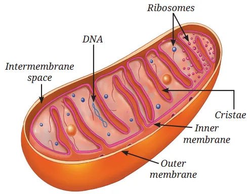
- They have two membranes.
- The inner membrane forms folds called cristae.
- Cristae increase the surface area for chemical reactions and facilitate energy production.
- Glucose and other molecules are broken down during cellular respiration.
- The released energy is stored as ATP (Adenosine Triphosphate).
- ATP acts as the energy currency of the cell.
6. Plastids
Plants contain special organelles called plastids, which are involved in food synthesis and storage.
Types of Plastids
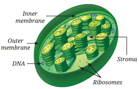
(a) Chloroplasts
- Green plastids.
- Contain the green pigment chlorophyll.
- Chlorophyll absorbs sunlight.
- They are the site of photosynthesis.
- They are double-membrane-bound organelles.
- The inner fluid is called the stroma.
- They contain their own DNA and ribosomes.
(b) Chromoplasts
- Contain pigments other than chlorophyll.
- May contain yellow, orange or red pigments.
- Give bright colours to flowers and fruits.
- These colours can help attract pollinators and fruit-eating animals.
(c) Leucoplasts
- Are colourless plastids.
- Store food materials such as:
- starch,
- oils, and
- proteins.
- For example, some leucoplasts in potato and taro store starch.
7. Vacuoles — Storage and Support
In Plant Cells
- A mature plant cell usually contains one large central vacuole.
- It is surrounded by a single selectively permeable membrane.
- It contains a watery fluid called cell sap.
- It stores:
- water,
- minerals,
- sugars, and
- waste materials.
- By storing water, it maintains pressure inside the cell and helps keep the plant cell firm.
- When the plant lacks water, the vacuole loses water, cells become less firm and the plant wilts.
In Animal Cells
- Vacuoles may sometimes be present.
- They are much smaller than plant vacuoles.
- They help in the temporary storage of materials.
Difference between Plant Cell and Animal Cell
| Basis | Plant Cell | Animal Cell |
|---|---|---|
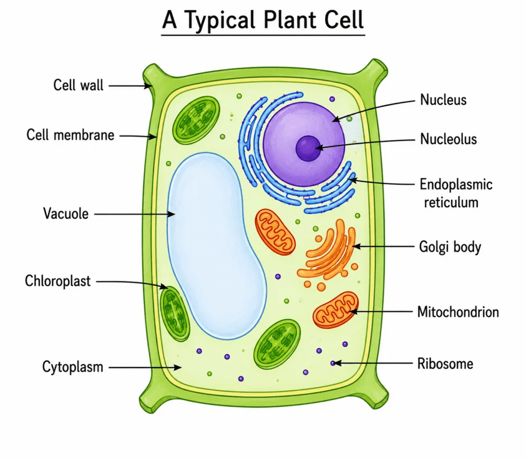
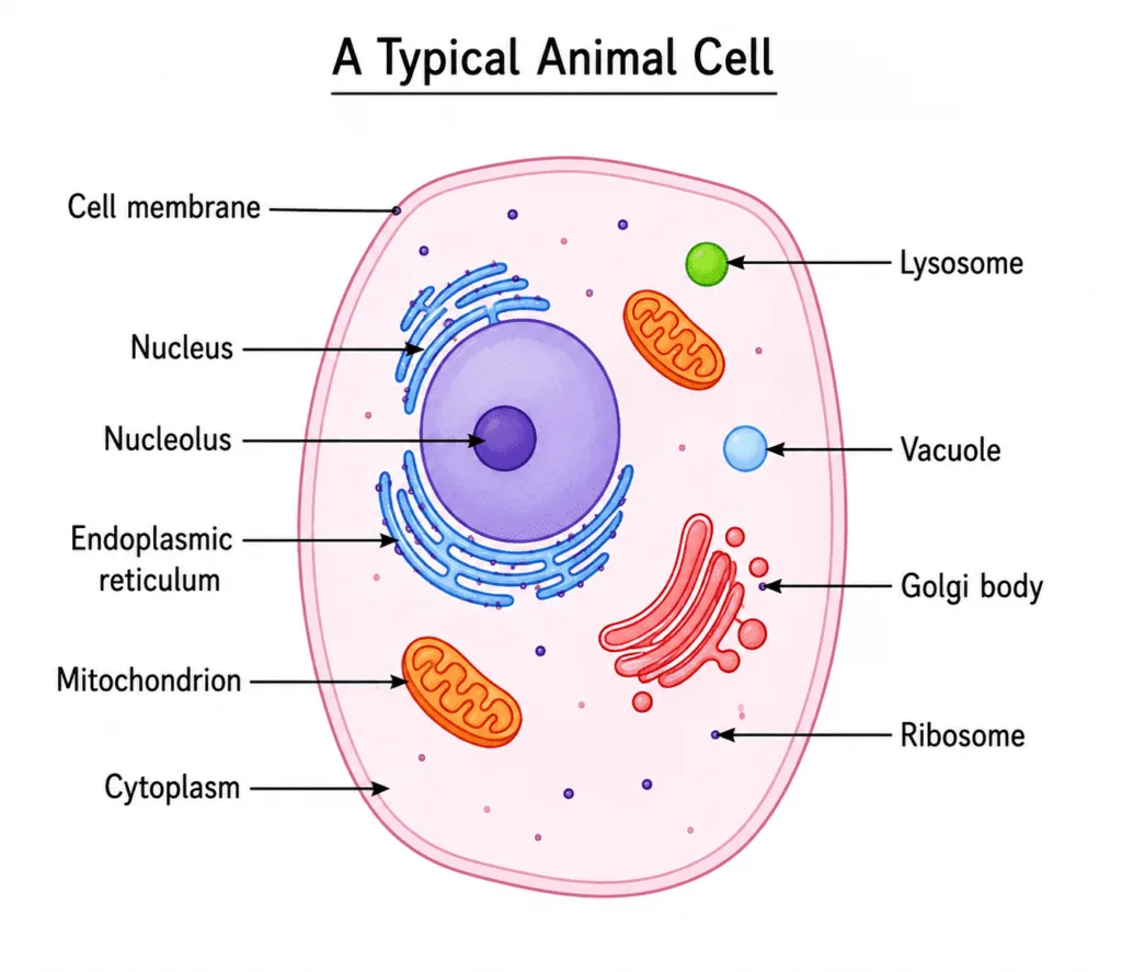
| 1. Cell wall | Present outside the cell membrane. It provides rigidity, support and helps maintain the shape of the cell. | Absent. Therefore, animal cells can change their shape more easily. |
| 2. Shape | Generally box-shaped and regularly arranged because of the rigid cell wall. | Generally irregularly arranged and more flexible because there is no cell wall. |
| 4. Plastids | Present. | Absent. |
| 5. Chloroplasts | Present in green plant cells; contain chlorophyll and help in photosynthesis. | Absent. |
| 6. Vacuoles | Usually has one large central vacuole containing cell sap. | Vacuoles may sometimes be present but are smaller than those in plant cells. |
| 7. Function of vacuole | Stores water, minerals, sugars and waste; helps maintain pressure and keeps the cell firm. | Helps in the temporary storage of materials. |
How do Normal Cells Grow and Divide?
Cells in our body can grow and divide to replace old, dead or damaged cells. Growth of an organism does not occur simply because individual cells become larger; cells can grow only up to a certain size, after which cell division produces new cells.
1. Cell Division
Cell division is the process by which new cells are formed from pre-existing cells.
Importance of Cell Division: Cell division helps organisms in:
- Growth
- Repair of damaged tissues
- Replacement of old or dead cells
- Reproduction
Types of Cell Division: There are two major types:
- Mitosis
- Meiosis
2. Mitosis
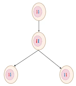
Mitosis is the most common type of cell division.
It increases the number of cells in the body. A human being begins life as a single fertilised egg, which divides repeatedly by mitosis to form the trillions of cells present in the body.
Main Features of Mitosis
- One parent cell divides.
- It produces two daughter cells.
- The daughter cells are genetically identical to the parent cell.
- Each daughter cell receives:
- the same DNA, and
- the same number of chromosomes as the parent cell.
- Thus, genetic information is largely maintained in body cells.
Functions of Mitosis
Mitosis is important for:
- Growth
- Repair
- Maintenance
- Replacement of cells
- Asexual reproduction
3. Meiosis
Meiosis is a type of cell division that produces gametes (sperm/egg) and occurs in the cells of reproductive organs.
Gametes are required for sexual reproduction.
In Animals/humans: Meiosis occurs in:
- Testes → produce sperm through meiosis.
- Ovaries → produce eggs through meiosis.
In Plants: Meiosis occurs in:
- Anthers → formation of pollen grains, which later produce sperm cells.
- Ovaries → formation of egg cells.
4. How Does Meiosis Occur?
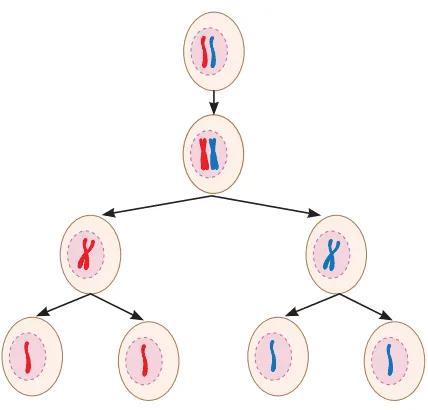
Meiosis involves two successive divisions.
First Division
- The parent cell divides into two daughter cells.
- The number of chromosomes in each daughter cell becomes half that of the parent cell.
Second Division
- Each of the two daughter cells divides again.
- This division is similar to mitosis.
- Finally, four daughter cells are formed.
- Each daughter cell has half the number of chromosomes of the original parent cell.
5. Why Is Meiosis Important?
- Meiosis produces gametes for sexual reproduction.
- The gametes produced during meiosis contribute to variation and diversity among organisms.
- Therefore:
- Children resemble their parents.
- But children are not exactly identical to their parents.
- The original chromosome number is restored when gametes from two individuals combine.
6. Mitosis vs Meiosis
| Basis | Mitosis | Meiosis |
|---|---|---|
| Main role | Growth, repair, maintenance and asexual reproduction | Sexual reproduction |
| Where it occurs | Body cells | Cells of reproductive organs |
| Number of divisions | One division | Two successive divisions |
| Daughter cells produced | 2 | 4 |
| Chromosome number | Same as parent cell | Half of parent cell |
| Genetic similarity | Daughter cells are genetically identical to the parent | Produces variation and diversity |
| Importance | Maintains genetic information in body cells | Produces gametes and creates genetic diversity |
7. What Happens When Cell Division Goes Wrong?
Errors in Mitosis: Errors in mitosis may lead to:
- Uncontrolled cell division
- Formation of tumours
- Abnormal chromosome numbers in body cells.
Errors in Meiosis: Errors in meiosis may result in:
- Genetic disorders
- Developmental problems
- Distinctive physical features
- Early pregnancy loss
- Reduced fertility.
Cell Theory
According to the classical Cell Theory:
- All living organisms are made up of one or more cells.
- The cell is the basic unit of structure and function in living beings.
- All cells arise from pre-existing cells.
Do Cells Grow and Reproduce Forever?
No. Cells do not grow and reproduce forever.
Cells:
- Grow in a controlled manner.
- Divide when required.
- Stay in their appropriate location.
- Perform their specialised functions.
- Eventually die when they are no longer needed.
- Dead cells are replaced by new cells performing the same function.
Therefore, every cell has a definite life span.
Contact Inhibition
In many animal cells, cell division usually stops when cells come into contact with neighbouring cells. This phenomenon is called contact inhibition.
Importance: Contact inhibition helps control the growth and division of cells.
What happens in cancer cells?
Cancer cells can lose contact inhibition. As a result keep dividing uncontrollably, leading to the
formation of tumours.
Plant cells grow differently from animal cells.
- Plant cells have rigid cell walls.
- Therefore, plant cells do not show contact inhibition in the same way as many animal cells.
- They follow a different pattern of growth.
Related posts:
 Matter in Our Surroundings Class 9 Important Questions
Is Matter Around Us Pure? Class 9 Important Questions
The Fundamental Unit of Life (Class 9) Important Questions
Science Class 9 Important Questions
Motion Class 9 Science Important Questions
Force and Laws of Motion Class 9 Important Questions
Gravitation Class 9 Important Questions
Matter in Our Surroundings Class 9 Important Questions
Is Matter Around Us Pure? Class 9 Important Questions
The Fundamental Unit of Life (Class 9) Important Questions
Science Class 9 Important Questions
Motion Class 9 Science Important Questions
Force and Laws of Motion Class 9 Important Questions
Gravitation Class 9 Important Questions
 Cell The Building Block of Life Class 9 Questions and Answers | NCERT Exploration Chapter 2